suPAR was first studied in a disease that recurs after transplant
Focal segmental glomerulosclerosis can return in a transplanted kidney within days. The new kidney is healthy, so whatever is destroying it arrived with the recipient.
The disease destroys podocytes, cells whose interlocking foot processes form the slits that plasma is filtered through. Primary focal segmental glomerulosclerosis returns in 30% to 80% of transplanted kidneys. In someone who has already lost one graft to recurrence, the risk for the next is 80% to 100% (Shoji, Nephron 2020). Something in the blood does this, and the field looked for it for decades without finding it.
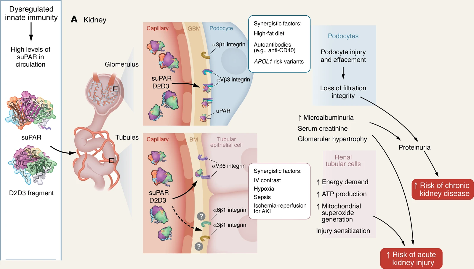
suPAR was proposed as that factor in 2011. It was raised in roughly two thirds of people with primary focal segmental glomerulosclerosis, highest before transplant in those whose disease recurred, and lowered by plasmapheresis. In mice, sustained suPAR expression produced protein in the urine and flattened the podocyte's foot processes. A mutant unable to bind αvβ3 integrin produced neither, and an anti-uPAR antibody prevented the injury (Wei, Nat Med 2011). That built on an earlier finding in mice that uPAR signaling in the podocyte itself activates αvβ3 integrin, producing foot process effacement and proteinuria (Wei, Nat Med 2008).
suPAR predicts kidney decline before standard tests show it
Whether suPAR causes that one rare disease is still argued. Its association with kidney disease in general is well established, and the evidence for it came from people who never had focal segmental glomerulosclerosis.
We measured suPAR in 3,683 participants of the Emory Cardiovascular Biobank and followed their kidney function. Filtration fell by 4.2 mL/min/1.73 m² per year in the highest quartile of suPAR, against 0.9 in the lowest. Among the 1,335 who did not have chronic kidney disease at the start, the highest quartile carried a multivariable-adjusted hazard ratio of 3.13 (95% CI 2.11–4.65) for developing it, against the lowest quartile. The steepest decline of all was in the 921 participants whose filtration was entirely normal to begin with, a group for whom standard testing gives no warning (Hayek, N Engl J Med 2015).
The finding has since held across different diseases and ancestries. Among 898 European children, five-year survival free of kidney failure was 64.5% in the lowest quartile of suPAR and 35.9% in the highest (Schaefer, JAMA Pediatr 2017). Among 649 adults with autosomal dominant polycystic kidney disease, 68% of the highest tertile reached stage 3 chronic kidney disease within three years, against 22% of the lowest (Hayek, J Am Soc Nephrol 2019). suPAR also predicts progression in 2,391 Chinese patients with chronic kidney disease (Lv, Nephrol Dial Transplant 2020). In Black Americans with chronic kidney disease, it predicts risk even after accounting for filtration measured directly rather than estimated (Luo, Clin J Am Soc Nephrol 2018).
suPAR is made by immune cells, not by the kidney
Immune cells make suPAR, and the kidney is one of the organs it reaches.
In mice, immature myeloid cells in the bone marrow produce the high levels seen in disease. Transferring those cells into healthy animals transfers the proteinuria (Hahm, Nat Med 2017). In people, bone marrow from patients with chronic kidney disease carries raised TNFα and suPAR alongside inflammatory monocytic cells. Myeloid cells reprogrammed by TNFα in culture secrete more suPAR alongside other cytokines; in experiments, that secretome disorganized the podocyte's cytoskeleton and impaired filtration in zebrafish (Spear, J Am Soc Nephrol 2026).
This is also why a suPAR level does not measure kidney function. The kidneys filter part of it, and they are not the only organ that removes it: regional blood sampling in people finds suPAR taken up across the heart as well as the kidney (Chew-Harris, Clin Biochem 2019). A dialysis session that removes 58% of β2-microglobulin leaves suPAR unchanged (Kampmann, Scand J Clin Lab Invest 2024). A level reflects the inflammatory state that produces it (Hayek, Circ Res 2026).
At the filter, suPAR arriving from the blood engages αvβ3 integrin together with RAGE on podocytes. In mouse podocytes, the pairing is required for the signaling that flattens foot processes (Kim, Biochim Biophys Acta Mol Basis Dis 2021). A second uPAR isoform forms a dimer; in mice, it signals through β3 integrin and c-Src to produce proteinuria and glomerulosclerosis, which a Src inhibitor reduced (Wei, J Clin Invest 2019). In D2D3-transgenic mice, the fragment produced kidney disease and depleted the insulin-producing beta cell, and an anti-uPAR antibody restored beta cell mass (Zhu, Sci Transl Med 2023).
APOL1 kidney risk depends on suPAR
Two variants in the APOL1 gene raise the risk of kidney failure sharply in people of recent African ancestry. Most carriers never develop kidney disease, so something else has to be present.
In two large, unrelated cohorts, the decline in kidney function associated with the APOL1 risk variants depended on the person's suPAR level. The genetic risk was attenuated when suPAR was low and strengthened when it was high. The risk proteins bind suPAR-activated αvβ3 integrin more tightly than the reference protein does. In mice, they caused proteinuria only when suPAR was present (Hayek, Nat Med 2017). The variants answer a signal the immune system is already sending.
In a separate cohort of Black Americans with chronic kidney disease, suPAR predicted worsening proteinuria only among those carrying two APOL1 risk alleles (Luo, Clin J Am Soc Nephrol 2018). A genetic risk that had looked fixed turns out to depend on a circulating protein, and circulating proteins can be lowered.
A level drawn before a procedure predicts acute kidney injury
Much of the kidney injury that happens in a hospital is scheduled: a contrast injection, a bypass run, an admission to intensive care. A level drawn beforehand identifies who is at risk.
We measured suPAR before coronary angiography in 3,827 patients, before cardiac surgery in 250, and on admission to intensive care in 692. Patients in the highest quartile had a multivariable-adjusted odds ratio of 2.66 (95% CI 1.77-3.99) for acute kidney injury within seven days, adjusted for demographic, clinical and procedural factors including baseline kidney function. In the same study, mice overexpressing suPAR had worse injury from contrast material, and human kidney tubular cells exposed to suPAR showed oxidative stress. An anti-uPAR antibody prevented both (Hayek, N Engl J Med 2020).
Among 352 patients admitted with COVID-19, acute kidney injury occurred in 6% of the lowest suPAR tertile and 46% of the highest. Nobody with a level below 4.60 ng/mL needed dialysis (Azam, J Am Soc Nephrol 2020). In sepsis, mice lacking suPAR were protected and mice overexpressing it did worse (Nusshag, JCI Insight 2023). Some of the proteinuria of a severe viral illness comes from the immune response itself, acting through suPAR on podocyte integrins. The evidence comes from African green monkeys infected with SARS-CoV-2 and from mice engineered to carry high suPAR (Wei, Nat Commun 2023).
Blocking suPAR works in mice; no human trial has reported
Every mechanism above has a matching intervention that works in an animal, but none has yet become a treatment for a person.
In mice, an anti-uPAR antibody prevents suPAR-driven proteinuria, reduces contrast-induced kidney injury and restores beta cell mass (Wei, Nat Med 2011; Hayek, N Engl J Med 2020; Zhu, Sci Transl Med 2023). Plasmapheresis lowers suPAR in people, but it removes much else besides, so any effect it has cannot be pinned on suPAR (Wei, Nat Med 2011). The one trial built around this biology gave rituximab to nine adults with treatment-resistant disease selected for a high suPAR, and proteinuria did not improve (Hladunewich, Kidney Int Rep 2022). Rituximab depletes B cells and does not lower suPAR, so no trial has yet reported what happens when patients with kidney disease are given a drug that acts on suPAR itself.
There is more reason to try now. Besides sorting patients by risk, suPAR has a receptor complex on the podocyte, a genetic interaction that explains who is susceptible, a source in the bone marrow, and an effect that can be blocked in an animal at three separate points. Because it also reaches organs beyond the kidney, there is a case for treating cardiovascular, kidney and metabolic disease as one problem rather than three (Reiser, J Clin Invest 2026; Hayek, Circ Res 2026).
Read next: