A high suPAR precedes symptoms by years
These cohorts enrolled adults without known cardiovascular disease, the population in which risk prediction guides prevention.
We measured suPAR in 5,406 participants of the Multi-Ethnic Study of Atherosclerosis, all free of known cardiovascular disease at enrollment, and followed them for a median of 15 years. Coronary artery calcium, a measure of atherosclerotic burden, progressed faster at higher suPAR: over a median of 2.5 years it rose 103% in participants below 2.0 ng/mL and 229% in those above 3.0 ng/mL. Incidence rates of cardiovascular events followed the same gradient, 2.8 per 1,000 person-years in the lowest category and 9.0 in the highest. Each doubling of suPAR was associated with a higher hazard of cardiovascular events after multivariable adjustment (HR 1.46, 95% CI 1.29–1.65), and further adjustment for kidney function did not attenuate the association (Hindy, J Clin Invest 2022).
The association first appeared in the general population. In 2,602 Danish adults of the MONICA10 study, a single baseline measurement predicted incident cardiovascular disease, type 2 diabetes, cancer and death over a median of 12.6 years, independently of C-reactive protein (Eugen-Olsen, J Intern Med 2010). suPAR also predicted cardiovascular events independently of the Framingham Risk Score, and together with C-reactive protein it reassigned people at intermediate Framingham risk to low- or high-risk categories (Lyngbæk, Int J Cardiol 2013). The association extends to cancer survivors: in 845 survivors of breast cancer in the UK Biobank, free of coronary artery disease and heart failure at baseline, suPAR measured by proteomics (Olink) was associated with cardiovascular death or incident coronary artery disease over 13 years, with an adjusted subdistribution hazard ratio of 3.08 for the highest quartile against the lowest (Yadalam, J Am Heart Assoc 2025). A marker that predicts cardiovascular events more than a decade ahead in asymptomatic adults either reflects a disease process already under way or contributes to it.
Human genetics and a mouse model point to a causal role
No cohort, however large, can distinguish a marker from a cause. Human genetics and a mouse model address the question.
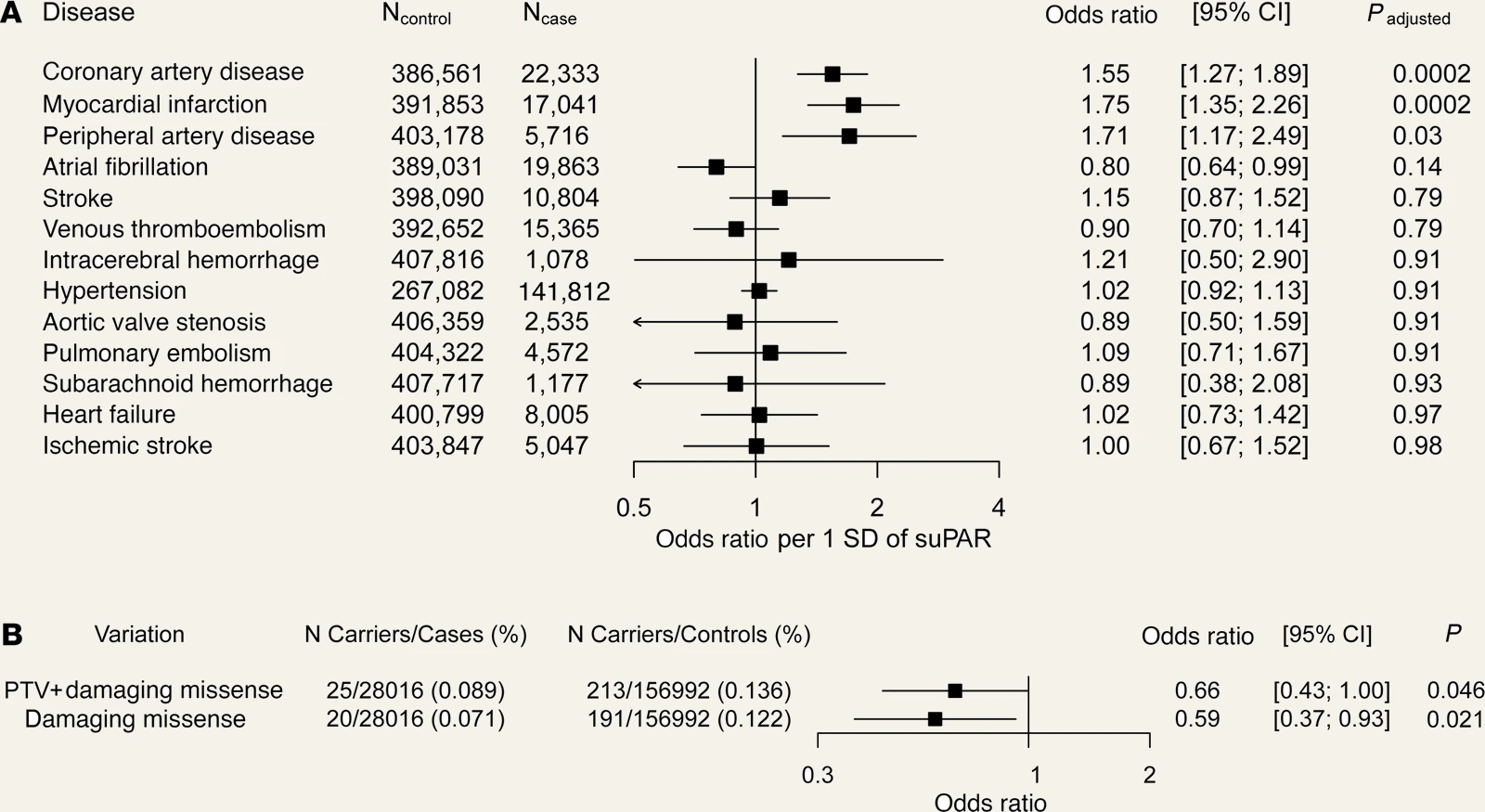
We performed a genome-wide association study of suPAR in 12,937 people across four cohorts and replicated the findings in 12,177 more. Two common missense variants in PLAUR, the gene encoding uPAR, were associated with suPAR levels; only one, rs4760, raised secreted suPAR when expressed in human embryonic kidney cells and in mice. Because alleles are allocated at random at conception, independently of lifestyle, rs4760 can serve as an instrument for Mendelian randomization. In 408,894 UK Biobank participants, genetically predicted suPAR was associated with coronary artery disease, myocardial infarction and peripheral artery disease, and with none of the ten other cardiovascular conditions tested (Hindy, J Clin Invest 2022).
Rare variants test the same question from the opposite direction. In a gene-level collapsing analysis of more than 280,000 UK Biobank exomes, carriers of rare damaging coding variants in PLAUR, expected to lower suPAR, had 41% lower odds of ischemic heart disease (95% CI 7%–63%) (Hindy, J Clin Invest 2022).
Transgenic mice overexpressing suPAR and wild-type C57BL/6J mice received the same Western diet and the same Pcsk9 gene transfer to induce atherosclerosis, and their cholesterol levels did not differ. The transgenic mice developed larger lesions: mean aortic root plaque volume was 1.55 mm³ against 0.90, necrotic core volume 0.18 mm³ against 0.05, and macrophage-positive area 47% of the plaque against 28% (Hindy, J Clin Invest 2022). In this model, raising suPAR alone was sufficient to worsen atherosclerosis. Circulating suPAR in these mice far exceeds any human concentration, so the experiment establishes the direction of the effect, not its size in humans.
Not another C-reactive protein
C-reactive protein is the inflammatory marker in clinical use, so a second marker has to add information to it. suPAR reflects a different process, and the two behave differently when the same questions are put to them.
C-reactive protein does not pass the same genetic test. Variants in its gene raise its concentration lifelong, by up to 30% per allele, yet show no association with coronary heart disease. In a Mendelian randomization analysis of 47 studies and 194,418 participants, the risk ratio per standard deviation of genetically raised C-reactive protein was 1.00 (95% CI 0.90–1.13) (Wensley, BMJ 2011). A general-population study of more than 40,000 people reached the same conclusion (Zacho, N Engl J Med 2008). Measured C-reactive protein is associated with events, but the genetic evidence indicates the protein itself is unlikely to be causal. The same approach applied to suPAR, with rs4760 as the instrument, gave an odds ratio of 1.55 per standard deviation for coronary artery disease. On the genetic evidence, C-reactive protein is a marker of coronary disease and suPAR a contributor to it.
C-reactive protein is an acute-phase reactant, synthesized by the liver in response to interleukin-6, so it tracks acute infection or tissue injury and falls as they resolve. suPAR is not an acute-phase reactant. Immune cells shed it from their own surface. Its level changes slowly: it shows no diurnal variation, has a within-individual coefficient of variation of about 10% (Thurison, Clin Chim Acta 2015), and is minimally affected by acute or short-term influences (Rasmussen, Front Immunol 2021). Smoking and SARS-CoV-2 infection change it quickly (Eugen-Olsen, Eur J Clin Invest 2016; Wei, Nat Commun 2023). In 296 patients with ST-elevation myocardial infarction treated with primary percutaneous coronary intervention, C-reactive protein rose and fell over the following days while suPAR remained stable. Over a median of 5.75 years, suPAR predicted all-cause mortality and recurrent myocardial infarction, and C-reactive protein predicted neither (Lyngbæk, Am J Cardiol 2012).
The two markers also identify different people. In 1,126 randomly sampled middle-aged adults, suPAR was associated with coronary artery calcium score and high-sensitivity C-reactive protein was not (Sørensen, Atherosclerosis 2014). In 1,703 patients with angiographically documented coronary artery disease, suPAR predicted cardiovascular death or myocardial infarction independently of C-reactive protein, high-sensitivity troponin, N-terminal pro-B-type natriuretic peptide and kidney function (Nikorowitsch, J Am Heart Assoc 2020). In 500 African Americans with type 2 diabetes, in a model containing suPAR, coronary artery calcium and high-sensitivity C-reactive protein, only suPAR remained associated with all-cause mortality (Hayek, J Am Heart Assoc 2018). Neither marker replaces the other, and a multi-biomarker risk score can include both (Desai, Am J Cardiol 2023).
We compared the two directly in BARI 2D, a randomized trial in patients with type 2 diabetes and coronary artery disease receiving intensive guideline-directed therapy. Both markers were measured in 2,277 participants at entry and in 1,978 again at one year. High-sensitivity C-reactive protein fell over that year, from a median of 2.07 to 1.30 mg/L, whereas no treatment arm lowered suPAR. Baseline suPAR was associated with the composite of death, myocardial infarction or stroke after multivariable adjustment (HR 1.40 per standard deviation, 95% CI 1.27–1.55). The association persisted after adjustment for C-reactive protein (HR 1.35, 95% CI 1.22–1.49). suPAR also stratified risk among patients whose C-reactive protein had normalized: across the four groups defined by both markers, five-year event rates ranged from 7.1% to 21.6% (Ismail, Diabetes Care 2026). A normal C-reactive protein on treatment does not exclude residual inflammatory risk.
In mice, persistently high suPAR primes the monocyte
Atherosclerotic lesions grow as circulating monocytes enter the arterial wall and become macrophages. In mice, sustained suPAR elevation primes these cells before they arrive, increasing their migration and amplifying their inflammatory response to a later stimulus.
In suPAR-transgenic mice without induced atherosclerosis, the aorta secreted more CCL2, a primary monocyte chemoattractant, and contained twice as many monocytes as in wild-type mice. Aortic monocytes expressed more CCR2, the receptor for CCL2, and circulating monocytes more CX3CR1. In a Transwell assay, more splenic monocytes from the transgenic mice migrated across the membrane, with or without added CCL2 (Hindy, J Clin Invest 2022).
Recombinant suPAR alone did not induce an inflammatory response in mouse bone marrow-derived macrophages, but it primed them. On stimulation with lipopolysaccharide and interferon-γ, primed macrophages produced more tumor necrosis factor-α, interleukin-1β and interleukin-6, and mounted larger NLRP3 responses, than unprimed ones. In suPAR-transgenic mice given a high-fat diet with L-NAME, a model of heart failure with preserved ejection fraction, sustained suPAR elevation expanded CCR2-positive inflammatory monocytes and macrophages in the heart, spleen and blood and worsened diastolic dysfunction (Singh, bioRxiv 2026).
On the monocyte surface, membrane uPAR localizes proteolysis and adhesion. Released into the circulation, suPAR engages αvβ3 integrin, RAGE and formyl peptide receptors on distant cells, including cells that never expressed uPAR, and primes monocytes toward chemotaxis and inflammatory activation (Hayek, Circ Res 2026).
In established disease, suPAR adds to the markers already in use
Most patients who need risk stratification already have established disease. In them, suPAR adds prognostic information to the markers already in use.
In people free of cardiovascular disease, median suPAR is about 2.5 ng/mL, and risk begins to separate above 3.0 (Hindy, J Clin Invest 2022). The median is 3.4 ng/mL in patients with heart failure (Hayek, J Card Fail 2023) and 4.7 ng/mL in patients with type 2 diabetes hospitalized for worsening heart failure (Ismail, J Card Fail 2026). Thresholds depend on sex. Women have levels about 10% higher than men after multivariable adjustment, and in patients with coronary artery disease the optimal cut-offs for mortality were 4.4 ng/mL in women and 3.2 in men; above them, mortality was similar in both sexes (Mehta, J Am Heart Assoc 2020). They also depend on the assay. Every suPAR concentration and cut-off on this page was measured with the same ELISA (suPARnostic), and values from other immunoassays or proteomic platforms cannot be read against them.
Among 1,116 patients with heart failure followed for a median of 6.2 years, each doubling of suPAR was associated with all-cause death after adjustment for clinical characteristics and B-type natriuretic peptide (HR 2.30, 95% CI 1.90–2.77). The association did not differ between reduced and preserved ejection fraction, and adding suPAR to a model containing B-type natriuretic peptide improved discrimination for death (C-statistic increase 0.027, 95% CI 0.009–0.045) (Hayek, J Card Fail 2023). suPAR also stratifies risk in heart failure with preserved ejection fraction, where treatment options are fewest. Among 406 such patients in the North American TOPCAT cohort, the five-year cumulative incidence of cardiovascular death, cardiac arrest or heart failure hospitalization was 44% in the highest suPAR tertile and 14% in the lowest, and the association was independent of natriuretic peptides (Hutten, ESC Heart Fail 2025).
The association extends beyond the coronary circulation. In 5,810 patients undergoing cardiac catheterization, suPAR was associated with prevalent peripheral arterial disease, one of the phenotypes the Mendelian randomization identified, and predicted death and peripheral arterial events (Samman, Atherosclerosis 2017). Reduced kidney function is associated with cardiovascular events independently of conventional risk factors (Go, N Engl J Med 2004). In 4,994 patients with chronic kidney disease, mediation analysis attributed about two thirds of suPAR's association with cardiovascular death to a direct effect rather than to kidney function (Sommerer, Kidney Int Rep 2023). This is consistent with kidney and cardiovascular disease sharing an upstream driver.
Current cardiovascular treatments do not lower suPAR
Three randomized trials have measured suPAR before and after treatment. No treatment lowered it relative to its comparator.
In BARI 2D, suPAR did not fall over one year under either glycemic strategy or either revascularization strategy. The other two trials tested heart failure drugs. Spironolactone did not change suPAR at one year in the North American TOPCAT cohort (Hutten, ESC Heart Fail 2025). Sotagliflozin reduced the SOLOIST-WHF primary composite of cardiovascular death and heart failure events by a third (HR 0.67, 95% CI 0.52–0.85). suPAR fell no more than with placebo, and the treatment effect was consistent across suPAR quartiles (Ismail, J Card Fail 2026).
Sotagliflozin's benefit is unlikely to operate through suPAR, and the risk suPAR marks persists in patients treated to target. Inflammation is a validated cardiovascular target: blocking interleukin-1β with canakinumab reduced recurrent cardiovascular events without lowering lipids (Ridker, N Engl J Med 2017). A pathway that current therapies do not reach is therefore a candidate drug target as well as a biomarker.
suPAR can be lowered by other means. In a randomized study of 48 smokers, four weeks of cessation lowered suPAR to levels no longer different from those of never-smokers, while C-reactive protein did not change (Eugen-Olsen, Eur J Clin Invest 2016). In a Danish population cohort, healthier diet and greater physical activity were associated with lower suPAR five years later (Haupt, Immun Ageing 2019).
No suPAR-blocking drug has yet been tested against a cardiovascular endpoint
No drug in clinical use lowers suPAR. The first antibodies built to block it are now being tested in patients.
A monoclonal antibody against uPAR attenuated contrast-induced acute kidney injury in suPAR-overexpressing mice (Hayek, N Engl J Med 2020). One antibody of that kind has completed a phase 1 safety study in healthy volunteers and is now in a randomized, placebo-controlled trial in patients with glomerular kidney disease (Yang, Front Endocrinol (Lausanne) 2026). None has yet been tested against atherosclerosis or any cardiovascular endpoint. Until one is, the evidence supports suPAR as a tool for risk stratification, not yet as a validated therapeutic target. Its immediate application is identifying patients with residual inflammatory risk whose glucose, blood pressure and lipids are already controlled.
Read next: