uPAR aims proteolysis at the cell–matrix interface
Leukocytes reach a site of inflammation by degrading the extracellular matrix in their path. uPAR lets them aim that proteolysis.
The urokinase plasminogen activator receptor sits on the outer face of the plasma membrane. It has no transmembrane or cytoplasmic domain and is held there by a glycosylphosphatidylinositol (GPI) anchor. Its three domains, D1, D2 and D3, form a concave shape around a central cone-shaped cavity, into which the receptor-binding amino-terminal fragment of urokinase inserts (Huai, Science 2006). The gene is PLAUR (Casey, Blood 1994), and a common missense variant in it raises circulating suPAR.
Urokinase converts plasminogen to plasmin, a protease that degrades extracellular matrix proteins. In solution that activity would be diffuse. Bound to uPAR, urokinase is concentrated at the interface between cell and matrix, so proteolysis occurs where the cell is migrating (Appella, J Biol Chem 1987).
uPAR also binds the matrix protein vitronectin with high affinity and independently of urokinase, which makes it an adhesion receptor in its own right (Wei, J Biol Chem 1994). It acts through lateral partners, including the integrin Mac-1 on monocytes, whose adhesive function it regulates (Simon, Blood 1996).
uPAR is induced when immune cells are activated
uPAR expression is inducible. The receptor is stored inside the cell, mobilized to the surface on activation, and upregulated to different degrees by different stimuli.
Mo3, an activation antigen of human monocytes, proved to be uPAR (Min, J Immunol 1992). Human neutrophils store the receptor in secretory vesicles and in a compartment close to the specific granules, and translocate it to the plasma membrane on stimulation (Plesner, Blood 1994). Surface expression can therefore rise within minutes without new protein synthesis. Over longer periods, interferon-γ and tumor necrosis factor-α increase the number of receptors on human monocytes (Kirchheimer, J Immunol 1988), and hypoxia increases uPAR messenger RNA and protein through a separate, heme protein-dependent pathway (Graham, Blood 1998).
Surface and soluble receptor are regulated independently. In U937 monocytic cells, interferon-γ raised surface uPAR 2.1-fold without increasing release. Tumor necrosis factor-α left surface levels unchanged but increased uPAR messenger RNA, doubled the intracellular pool and raised release into the medium 2.5-fold (Sitrin, Blood 1994). A rising blood level can therefore reflect increased shedding with no change in surface expression.
Because uPAR is induced on demand, its circulating level is informative. The level of a housekeeping protein, expressed constitutively, says little about a person. A protein released on immune activation reflects how often, and how strongly, that activation has occurred.
Leukocyte migration depends on uPAR
uPAR is required for leukocyte migration, in human cells in vitro and in mice.
In human monocytes, an anti-uPAR monoclonal antibody reduced chemotaxis toward the formyl peptide fMLP to 14.2% of that in untreated cells, comparable with random migration. A neutralizing antibody against urokinase had no significant effect (88.2% of untreated cells) (Gyetko, J Clin Invest 1994). Migration therefore depends on the receptor rather than on the catalytic activity of the urokinase it binds.
uPAR-deficient mice develop normally and are fertile (Bugge, J Biol Chem 1995). In thioglycollate-induced peritonitis, however, they recruit about 50% fewer leukocytes to the peritoneum, and their granulocytes almost entirely fail to migrate (May, J Exp Med 1998). In Pseudomonas aeruginosa pneumonia, their neutrophil recruitment to the lung is markedly reduced and bacterial clearance is impaired, whereas urokinase-deficient mice recruit neutrophils as well as wild-type mice (Gyetko, J Immunol 2000). No inherited uPAR deficiency has been described in humans, so whether these phenotypes apply to people is unknown.
suPAR is released by two routes that yield different molecules
suPAR is the receptor freed from its anchor. It is released by two routes that yield different molecules.
Cleavage of the GPI anchor releases the intact three-domain receptor into the circulation. Alternatively, urokinase itself or plasmin cleaves the linker between D1 and D2, which releases free D1 and a two-domain fragment (Høyer-Hansen, J Biol Chem 1992; Sidenius, FEBS Lett 2000).
The second route changes the receptor's function. Removing D1 abolishes uPAR's lateral interaction with integrins and its regulation of integrin-dependent adhesion (Montuori, J Biol Chem 2002). The cut also exposes a linker sequence that binds and activates FPRL1, a G protein-coupled chemotactic receptor, so the soluble two-domain fragment becomes a chemoattractant for monocytes (Resnati, Proc Natl Acad Sci U S A 2002). Activated human neutrophils release this fragment rapidly (Pliyev, Mol Cell Biochem 2009).
A measured suPAR concentration therefore combines forms with different biology, and assays whose antibodies recognize different domains capture different mixtures of them.
suPAR signals to cells that did not make it
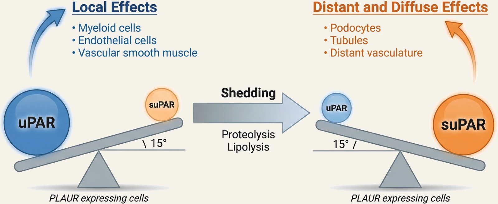
Released from the cell, suPAR competes with the anchored receptor for urokinase and activates signaling in cells elsewhere in the body. When that signaling is sustained, it can lead to disease in the target organ.
In solution the receptor still binds urokinase, but no longer localizes it. Recombinant soluble uPAR scavenges urokinase, blocking its binding to cells, and attenuates plasminogen activation (Wilhelm, FEBS Lett 1994; Behrendt, FEBS Lett 1996). In paroxysmal nocturnal hemoglobinuria, where plasma suPAR is elevated, the excess competes with membrane uPAR on normal neutrophils for urokinase and inhibits cell-associated fibrinolytic activity (Ninomiya, Int J Hematol 1997).
The binding surfaces keep working after the anchor goes. In cultured mouse podocytes, suPAR engages αvβ3 integrin in a complex with the receptor for advanced glycation end-products (RAGE), which activates Src and Rac1 signaling. Serum from a patient with recurrent focal segmental glomerulosclerosis increased Src phosphorylation in these cells, and a suPAR-neutralizing antibody abolished the effect (Kim, Biochim Biophys Acta Mol Basis Dis 2021).
Membrane uPAR acts on the cell that made it, where it localizes proteolysis and adhesion. Soluble suPAR signals to cells that did not make it. Our 2026 review describes these as the local and diffuse modes of one gene product, with distinct biology (Hayek, Circ Res 2026).
A suPAR level can only be compared on the same assay
Immunoassays and proteomic platforms both report a value called suPAR, but on different scales. The assay determines whether a level can be compared with a published threshold.
The reference method is the sandwich enzyme-linked immunosorbent assay (ELISA), which reports an absolute concentration. The two ELISAs in common use disagree: in the same samples, the suPARnostic and Quantikine assays had a Spearman correlation of 0.75, and suPARnostic values ran on average 50% higher (Vasbinder, J Nephrol 2023). A threshold therefore applies only to the assay on which it was derived.
The other route is high-throughput proteomics, in which suPAR is one of thousands of proteins measured on an aptamer (SomaScan) or proximity extension (Olink) platform. These platforms report relative abundance, normalized within a batch, rather than a concentration. In the Jackson Heart Study, SomaScan correlated with the Quantikine ELISA at a Spearman coefficient of 0.29, and with suPAR as the only predictor, Harrell's C-statistic for cardiovascular death was 0.74 for the ELISA and 0.57 for SomaScan (Vasbinder, J Nephrol 2023). Proteomics ranks risk within a cohort, but its values cannot be read against a published cut-off. An individual patient's level has to come from an ELISA.
In health, suPAR sits at a slow-moving set-point
A healthy person carries suPAR at a set-point that varies with age, sex and lifestyle.
suPAR has been measurable in blood since an ELISA was described in 1995 (Mizukami, Blood 1995; Brünner, APMIS 1999), and levels in healthy adults run between roughly 2 and 4 ng/mL: a median of 1.9 ng/mL in never-smokers in one study (Eugen-Olsen, Eur J Clin Invest 2016), and 4.0 ng/mL in an older Danish general-population cohort that included smokers (Eugen-Olsen, J Intern Med 2010). No single reference range applies: the value depends on the population and on the assay.
In 5,538 participants of Inter99, a Danish general-population study, suPAR was higher with older age, in women and with lower socioeconomic status. After multivariable adjustment, the largest differences, at least 1 ng/mL, were with daily heavy smoking and morbid obesity (body mass index ≥40 kg/m²) (Haupt, Biomark Insights 2014). Levels also change within individuals. In a randomized trial, smokers who quit saw suPAR fall by a mean of 1 ng/mL (about 30%) within four weeks, to the level of never-smokers (Eugen-Olsen, Eur J Clin Invest 2016). An unhealthy diet and low physical activity were associated with higher levels five years later (Haupt, Immun Ageing 2019).
suPAR is useful because it is not an acute-phase reactant. C-reactive protein and the other hepatic acute-phase proteins rise within hours of an infection, surgery or myocardial infarction and fall as the insult resolves, whereas suPAR is minimally affected by such acute changes (Rasmussen, Front Immunol 2021). It reflects slower processes, closer to cumulative immune activation, so a single measurement in a healthy person predicts kidney and cardiovascular outcomes years later (Thunø, Dis Markers 2009).
Two exposures change it quickly: smoking, as the cessation trial above shows, and SARS-CoV-2 infection, which raises suPAR in patients and in experimentally infected African green monkeys (Wei, Nat Commun 2023). suPAR is also elevated in HIV infection, but unlike the other biomarkers measured, it did not fall during the first year of suppressive antiretroviral therapy (Hoenigl, Clin Infect Dis 2019).
suPAR is implicated in cardiovascular, kidney and metabolic disease
Elevated suPAR is associated with adverse outcomes across a wide range of human disease. Working alongside other groups, we have spent a decade asking why.
Clinical, genetic and experimental evidence now implicates suPAR in the pathogenesis of cardiovascular, kidney and metabolic disease. The genetic evidence rests on rs4760, a common missense variant in PLAUR that raises circulating suPAR. In Mendelian randomization in the UK Biobank, genetically predicted suPAR was associated with atherosclerotic disease (OR for coronary artery disease 1.55 per SD) (Hindy, J Clin Invest 2022). The three conditions cluster: nearly 90% of US adults have stage 1 or higher cardiovascular-kidney-metabolic syndrome, and all-cause mortality rises with each stage (HR 2.87, 95% CI 1.98–4.17, stage 4 against stage 0) (Cheema, Am Heart J Plus 2026). Traditional risk factors explain this clustering incompletely: patients with well-controlled glucose, lipids and blood pressure still progress, which implies additional mediators between systemic inflammation and end-organ disease (Hayek, Circ Res 2026). suPAR may be one of them.
Where the lab has followed it:
- suPAR and kidney disease Prediction of chronic kidney disease before glomerular filtration declines, the interaction with APOL1 risk variants, and suPAR signaling in podocytes.
- suPAR and cardiovascular disease Human genetics, Mendelian randomization and mouse models placing suPAR upstream of atherosclerosis.